
Contact information
_______________________
5th floor office, room B501a
Tel: +358 2941 25269
1st floor office, room B117a
Tel: +358 2941 25271
Biomedicum Helsinki
PO Box 63 (Haartmaninkatu 8)
FI-00014 University of Helsinki
Finland
| 
|
Instruments
We recommend that you check the excitation and emission spectrum of your fluorochromes prior to the use of the equipment. Note that with the confocal workstation your experimental restrictions are largely based on the laser excitation lines, and with the wide-field workstation on the available emission filter combinations.
Widefield microscopes
![EVOS FL]() |
ThermoFisher Scientific EVOS FL (Room B501b)
Inverted epifluorescence microscope with long working distance objectives for bright-field, phase contrast, and fluorescence microscopy. Mainly used for cells growing on plastic bottom dishes. Image acquisition through integrated camera and software, images stored directly to memorystick. |
 |
Zeiss Axio Imager (1 & 2) (Room B501b)
Upright epifluorescence microscope.Image acquisition through Hamamatsu Orca Flash 4.0 LT camera and Zen software. Motorized stage for mosaic imaging. |
 |
Zeiss Axio Imager (3) with ApoTome (Room B502b)
Upright epifluorescence microscope. Equipped with a Zeiss ApoTome for optical sectioning with structured illumumination. Image acquisition through Hamamatsu Orca R2 camera and Zen software. Motorized stage for mosaic imaging. |
Confocal microscopes
Live cell widefield microscopes
 |
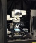
Nikon Eclipse Ti-E N-STORM imaging system (Room B115a2)
Nikon Eclipse Ti-E inverted widefield microscope with a 4 laser illumination unit for STORM super-resolution and TIRF imaging. The system has 405, Argon, 561 and 647 laserlines and is equiped with a Nikon PFS (Perfect focus system) for live cell imaging. |
 |
Nikon Eclipse Ti-E imaging system (Room B115a1)
Nikon Eclipse Ti-E inverted widefield microscope with a Lumencor Spectra X light engine for fast, triggered imaging and a Nikon PFS3 (Perfect focus system) for live cell imaging. |
Multiphoton microscopes
 |
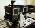
Zeiss 7 MP with OPO (Room AP29b)
Upright multiphoton microscope optimized for intravital imaging of anesthetized small laboratory animals. The system allows acute as well chronic in vivo imaging experiments on internal (e.g. brain, kidney, liver etc.) and external organs (e.g. skin and eye). The system can also be used for imaging of fixed thick tissue samples.
|
 |
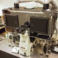
Leica TCS SP8 MP CARS confocal (Room B116a)
|
In vivo and whole mount imaging
 |
PerkinElmer IVIS 100 imaging system (Room AP29a)
Bioluminescense and fluorescence imaging of live animals. The imaging chamber has room for up to five mice or two rats. Peltier cooled, back-thinned, back-illuminated CCD camera for image acquisition and Living Image 2.20 software. Equipped with a gas anesthesia system. |
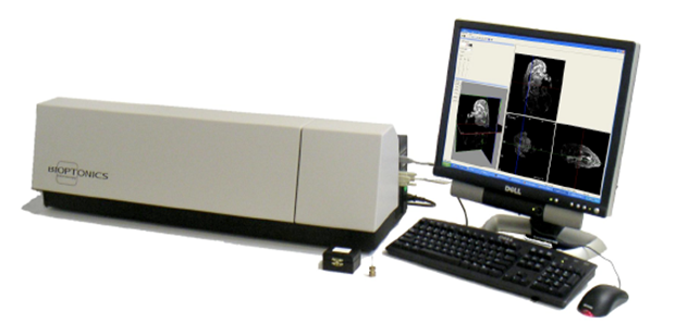 |
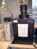
Optical Projection Tomography (OPT) (Room 504b)
The Bioptonics OPT 3001M Scanner is an optical projection tomography platform that produces high-resolution (15-30 microns) 3-Dimensional image reconstructions of both fluorescently stained and non-fluorescent specimens ranging in size from 1mm to 15mm. Applications include visualization of anatomy (phenotyping), gene expression (in situ hybridization), protein distribution (immunohistochemistry or GFP expression), transgenic visualization (LacZ). Typical specimens include mouse and chicken embryos, mouse/rat tissue and organs, zebrafish (5 days and older), drosophila, plants etc. |
High Content Screening
 |
Thermo Scientific CellInsight (Room B502b)
Cell-based high-content screening system for automated fluorescence image aqcuisition and quantitative analysis. Ideal for both RNAi and chemical compound screens. Up to four different fluorochromes can be used.
With CellInsight, you can study e.g. biochemical targets, signaling pathways, organelle status, phenotypic changes, and cell functions. Special bioapplications are available, for example cell cycle, motility, spreading, and viability analysis, as well as for molecular translocation, GPCR signaling, neurite outgrowth, and cytotoxicity. |
Computer workstations
 |
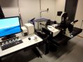
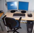
Imaging workstation 1 (Room B501 lobby)
HPZ840 Windows 7 workstation with 64 GB RAM and NVidia Quadro K2200 graphics card. General use workstation for image processing and analysis (also in 3D and 4D). HP ScanJet G4010 scanner for paper, film, and gels. |
 |
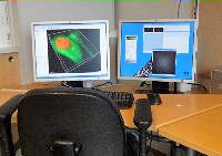
Imaging workstation 2 (Room B501 lobby)
Osborne with Intel Core2 Duo 3.0 GHz CPU, 4 GB RAM and dual monitors. Workstation for disk-based (virtual) rescanning and analysis of high-content screening data using Cellomics DiscoveryToolbox software. Spotfire DecisionSite 3D data visualization software; ImageProPlus for image processing and analysis; Living Image for analysis of IVIS data. IgorPro for graphing as well as data and image analysis. Also other imaging software. |
 |
Imaging workstation 3 (Room B501 lobby)
HP Z840 Windows 10 workstation with 256 GB RAM and NVidia Quadro M4000 graphics card. SVI Huygens Professional deconvolution with confocal, wide-field and time modules. Bitplane Imaris volume rendering software with measurement and co-localization modules. |
For Translational Cancer Biology and Genome-Scale Biology Programs internal use only
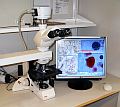 |
Leica DM LB
Research microscope for bright-field microscopy. Image acquisition through Olympus digital camera and Studio Lite software. |
Page updated
30.10.2017
Mail to Webmaster |